CO034 | VALIDATION OF A MICROFLUIDIC PLATFORM FOR HEMOSTASIS EVALUATION UNDER FLOW CONDITIONS: FROM PATHOGENETIC MECHANISMS TO THERAPEUTIC MONITORING
F. Boggio1|2, L. Padovan1|2, S. La Marca1, G. Donadoni1, A. Lecchi1|2, A. Artoni1, S. Bozzi3, A. Redaelli3, F. Peyvandi1|2 | 1Centro Emofilia e Trombosi A. Bianchi Bonomi, IRCCS Fondazione Ca’ Granda Ospedale Maggiore Policlinico di Milano, Italy; 2Dipartimento di Fisiopatologia Medico-Chirurgica e dei Trapianti, Università degli Studi di Milano, Italy; 3Dipartimento di Elettronica, Informazione e Bioingegneria, Politecnico di Milano, Italy
All claims expressed in this article are solely those of the authors and do not necessarily represent those of their affiliated organizations, or those of the publisher, the editors and the reviewers. Any product that may be evaluated in this article or claim that may be made by its manufacturer is not guaranteed or endorsed by the publisher.
Authors
Background and Aims. Conventional static assays for primary hemostasis evaluation fail to incorporate the crucial role of vascular hemodynamics. The development of strictly controlled in vitro systems capable of replicating physiological and pathological shear stress is required to overcome these limitations and accurately investigate congenital and acquired platelet disorders. The aim of the current study is to develop, validate on a healthy cohort, and preliminarily evaluate a microfluidic assay designed for the functional study of hemostasis, the dissection of pathogenetic mechanisms, and potential ex vivo therapeutic monitoring.
Methods. A microfluidic system was engineered for whole blood perfusion over functionalized surfaces to simulate venous and arterial shear rates via fluorescence microscopy. Specifically, microchannels were coated with collagen at two different concentrations (10 and 100 μg/mL) and blood was perfused at shear rates of 300 s⁻¹ and 1500 s⁻¹. Method validation was performed on 40 healthy donors to establish reference ranges, quantifying parameters such as fluorescence intensity, surface coverage, thrombi area, and number, with a customized MATLAB algorithm. The platform was subsequently applied to whole blood samples from patients with already known hemostatic defects: Glanzmann Thrombasthenia (GT), Bernard-Soulier Syndrome (BSS), delta-storage pool deficiency (delta-SPD), and Thrombotic Thrombocytopenic Purpura (TTP).
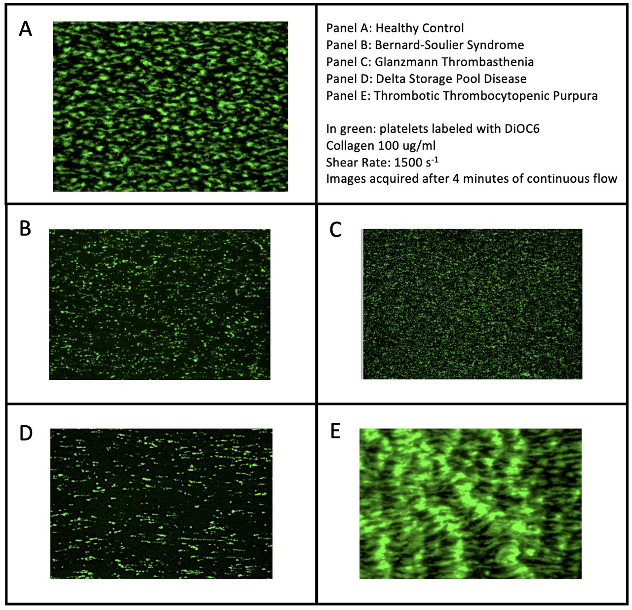
Results. Validation in the 40 healthy subjects demonstrated high intra- and inter-assay reproducibility, with an intraclass correlation coefficient >0.75 in most of the measured parameters, establishing stable quantitative profiles for platelet adhesion and aggregation. In preliminary testing, the device successfully discriminated pathological phenotypes under specific flow conditions (Figure 1, Panels A-E: representative fluorescence microscopy images of healthy control vs. pathological samples). Specifically, GT samples showed preserved primary adhesion to the substrate but a complete absence of three-dimensional thrombus formation. In contrast, BSS samples exhibited a severe defect in primary adhesion, which was markedly exacerbated at the higher shear rate of 1500 s⁻¹. Samples from delta-SPD patients displayed normal initial adhesion followed by impaired stability and early disaggregation of the secondary thrombus. On the contrary, patients with TTP showed severe platelet hyper-reactivity, resulting in massive microthrombi formation predominantly at 1500 s⁻¹.
Conclusions. The proposed microfluidic assay constitutes a translational tool for the pathophysiological investigation of primary hemostasis and provides a rationale for its use in pharmacological monitoring. However, methodological limitations persist: the assay requires qualified personnel and demands rigorous pre-analytical standardization.
Downloads
Citations




How to Cite

This work is licensed under a Creative Commons Attribution-NonCommercial 4.0 International License.