
CO-31 | WHEN NECK PAIN HIDES A SPINAL BLEED: ANTERIOR CERVICAL EPIDURAL HEMATOMA IN A CHILD WITH HEMOPHILIA B
E. Dalla Porta, C. Simion, E. Zanon, B. Stocco, L. Spiezia, P. Simioni | Hemophilia Center, University Hospital of Padua, Italy
All claims expressed in this article are solely those of the authors and do not necessarily represent those of their affiliated organizations, or those of the publisher, the editors and the reviewers. Any product that may be evaluated in this article or claim that may be made by its manufacturer is not guaranteed or endorsed by the publisher.
Authors
Introduction. Cervical anterior spinal epidural hematoma is an exceptionally rare bleeding manifestation in pediatric hemophilia, with only isolated cases reported in the literature. Prompt recognition is crucial to prevent irreversible neurological sequelae. We report a child with moderate hemophilia B who developed a cervical anterior epidural hematoma successfully managed with conservative treatment.
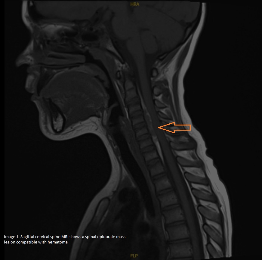
Case Report. A 9-year-old boy with moderate hemophilia B (factor IX activity 2–5.6%), diagnosed during infancy and receiving regular factor IX replacement therapy, presented with left shoulder pain following a prolonged car journey. Within 24 hours, cervical and dorsal pain developed without neurological deficits. Magnetic resonance imaging (MRI) revealed an anterior cervical epidural hematoma extending from C3 to C7, with a maximum thickness of 5 mm and a craniocaudal extension of approximately 55 mm, causing mild spinal cord compression (Image 1). Laboratory evaluation excluded the presence of factor IX inhibitors. Given the absence of neurological impairment and the limited degree of canal compromise, neurosurgical intervention was not indicated. Recombinant factor IX replacement therapy with nonacog-alfa was intensified to 1000 IU (approximately 35 IU/kg) every other day for two weeks, under close clinical and radiological monitoring. Follow-up MRI demonstrated marked regression of the hematoma, which became confined to the anterior epidural space between C4 and C6, with reduction of both thickness (1.5 mm) and longitudinal extension (28 mm). The patient became completely asymptomatic and experienced no further bleeding complications during follow-up.
Conclusions. Cervical epidural hematoma represents an uncommon and potentially life-threatening bleeding manifestation in children with hemophilia. This case highlights the importance of maintaining a high index of suspicion when persistent cervical, dorsal, or radicular pain occurs, even in the absence of neurological deficits. Early MRI assessment was crucial for diagnosis and therapeutic decision-making. Conservative management with prompt factor IX replacement therapy allowed complete clinical recovery and substantial radiological improvement, avoiding neurosurgical intervention. The unusual cervical anterior localization and favorable outcome underline the importance of rapid recognition and multidisciplinary management of atypical bleeding presentations in hemophilia.
Downloads
Citations




How to Cite

This work is licensed under a Creative Commons Attribution-NonCommercial 4.0 International License.