
CO-29 | A RARE BLEEDING PRESENTATION IN MODERATE HEMOPHILIA A: PARAVESICAL HEMATOMA WITH PENOSCROTAL EXTENSION MIMICKING A UROLOGICAL EMERGENCY
B. Stocco, C. Simion, E. Zanon, E. Dalla Porta, P. Simioni | Hemophilia Center, University Hospital of Padua, Italy
All claims expressed in this article are solely those of the authors and do not necessarily represent those of their affiliated organizations, or those of the publisher, the editors and the reviewers. Any product that may be evaluated in this article or claim that may be made by its manufacturer is not guaranteed or endorsed by the publisher.
Authors
Background. Bleeding episodes in hemophilia typically involve joints and muscles, whereas deep pelvic hematomas are uncommon and may present with nonspecific symptoms, leading to diagnostic uncertainty. Paravesical hematomas are particularly rare and can mimic acute surgical or urological conditions. We report an unusual case of paravesical hematoma with penoscrotal extension in a patient with moderate hemophilia A.
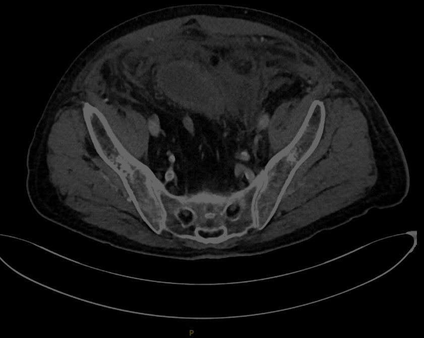
Case Report. An 81-year-old man with moderate hemophilia A (factor [F]VIII 3.6%), treated on demand with plasma-derived FVIII concentrate, presented with progressive lower abdominal pain, dysuria, nausea, bilious vomiting, and acute penile and scrotal swelling with ecchymosis. One month earlier, he had undergone abdominal wall reconstruction for an incisional hernia. Laboratory investigations showed leukocytosis, elevated C-reactive protein, and mild anemia (hemoglobin 11.2 g/dL). Ultrasound revealed a heterogeneous left paravesical collection measuring 8.8 × 5.6 × 3.8 cm without Doppler vascularization. Contrast-enhanced computed tomography confirmed a recent non-active paravesical hematoma measuring 10.3 × 9.5 × 5.3 cm, compressing and displacing the urinary bladder, with extension along the spermatic cord and into the penoscrotal region (Image 1). The patient was managed conservatively with factor VIII replacement therapy (37000 UI overall, for a period of ten days, starting from 70 UI/kg/day) and close clinical and radiological monitoring. No active bleeding was identified, and neither transfusion support nor surgical or radiological intervention was required. Hemoglobin reached a nadir of 9.1 g/dL during hospitalization before progressively increasing to 12.5 g/dL at discharge. Follow-up ultrasound demonstrated hematoma reduction to 7.3 × 5.9 × 3.5 cm with progressive organization of the collection. Clinical symptoms gradually resolved. Post-discharge magnetic resonance imaging showed slight reduction and resorption, confirming progressive spontaneous resolution.
Conclusions. This case expands the spectrum of atypical bleeding sites in hemophilia A and highlights paravesical hematoma as a rare cause of abdominal pain, urinary symptoms, and penoscrotal swelling. Clinicians should maintain a high index of suspicion for deep pelvic bleeding when these manifestations occur, even in patients with moderate disease. Early recognition and appropriate replacement therapy may result in favorable outcomes without the need for surgical or radiological intervention.
Downloads
Citations




How to Cite

This work is licensed under a Creative Commons Attribution-NonCommercial 4.0 International License.