PO06 | Extracellular vesicles profiles in patients with porto-sinusoidal vascular disease
S. Toffanin1, E. Campello1, A. Zanetto2, C. M. Radu1, E. Pinto2, K. Fernande2, C. Bulato1, B. Serena1, L. Spiezia1, P. Burra2, M. Senzolo2, P. Simioni1 | 1First Chair of Internal Medicine, Department of Medicine, University Hospital of Padua Medical School, Padua; 2Department of Surgery Oncology and Gastroenterology, Gastroenterology and Multivisceral Transplant Unit, University Hospital of Padua, Italy
All claims expressed in this article are solely those of the authors and do not necessarily represent those of their affiliated organizations, or those of the publisher, the editors and the reviewers. Any product that may be evaluated in this article or claim that may be made by its manufacturer is not guaranteed or endorsed by the publisher.
Authors
Background and Aims: Porto-sinusoidal vascular disorder (PSVD) has recently been proposed to delineate a group of hepatic vascular diseases characterized by lesions involving the portal venules and sinusoids, irrespective of the presence/absence of portal hypertension. Although data is still limited, several hypotheses and emerging evidence suggest that extracellular vesicles (EVs) might exert a functional role in the pathogenesis of PSVD. The analysis of EVs in PSVD may unveil disease-specific alterations in intercellular communication, contributing to understand the prothrombotic mechanisms and to discover novel biomarkers for diagnosis and prognosis. The study aims: i) to compare plasma-derived EVs in PSVD patients vs. healthy controls by flow cytometry; ii) to evaluate their potential involvement in disease progression.
Methods: Twenty-nine PSVD patients (median age 58 yrs; 20 males, 9 females) were included and compared with 9 (4 males and 5 females) aged matched (±3 yrs) healthy controls. Large extracellular vesicles (L-EVs) were isolated from platelet-poor plasma by centrifugation at 14,000 g for 30 min at 4°C, immunolabeled with calcein-AM, annexin V, CD41, CD62P, CD45, CD14, CD62E, anti-human-tissue factor (TF), CD105 and CD147. For EV size calibration, fluorescent polystyrene beads Gigamix were used to set a gate between 0.2 and 0.9 μm bead populations, defined as L-EVs gate. EVs were expressed as events/μl (absolute count) with the volume measurement of the CytoFLEX S.
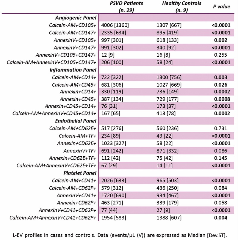
Results: All enrolled patients had histologically confirmed PSVD. Among them, 12 had unprovoked PSVD, 4 were drug-related, 3 were associated with gastrointestinal diseases, 3 with immune-mediated conditions, and 7 cases were linked to myeloproliferative disorders. Endothelium-derived L-EVs co-expressing calcein-AM, annexin V, CD62E, and TF were significantly increased in PSVD patients compared to healthy controls (p<0.0001), reflecting endothelial activation and prothrombotic predisposition (Table 1). Patients also exhibited a marked increase in platelet-derived L-EVs, particularly calcein-AM+/annexin V+/CD41+/CD62P+ L-EVs (p=0.004), indicating enhanced platelet activation and a contribution to vascular and coagulative responses (Table 1). Notably, endothelial and platelet L-EV significantly correlated with PSVD etiology (r=0.38, p=0.04), with patients affected by myeloproliferative disease–related PSVD showing significantly higher levels than those with other etiologies (p=0.046 and 0.032, respectively). Angiogenesis-related calcein-AM+/annexin V+/CD105+/CD147+ L-EVs were significantly elevated in patients compared to controls (p<0.0001), suggesting active vascular remodeling. Regarding the inflammatory panel, patients showed significantly lower levels of calcein-AM+/annexin V+/CD45+/CD14+ L-EVs compared to controls (p=0.0002), indicating reduced monocyte/leukocyte-driven inflammatory activation (Table 1).
Conclusions: In conclusion, patients with PSVD display a L-EV profile characterized by vascular remodeling, endothelial and platelet activation. On the other hand, patients exhibited significantly lower levels of inflammatory L-EVs, suggesting limited systemic immune activation. Our preliminary findings support the hypothesis that PSVD is primarily driven by endothelial dysfunction and platelet activation, rather than by leukocyte-mediated inflammation. The etiology of PSVD appears to influence L-EV levels.
Downloads
Citations


How to Cite

This work is licensed under a Creative Commons Attribution-NonCommercial 4.0 International License.