CO24 | Extracellular vesicles profiles in patients with wild-type transthyretin amyloidosis
S. Toffanin1, L. Spiezia1, C.M. Radu1, A. Napolitano1, A. Benetti1, C. Samà1, E. Campello1, L. De Michieli2, G. Sinigiani2, A. Cipriani2, P. Simioni1 | 1First Chair of Internal Medicine, Department of Medicine, University-Hospital of Padua Medical School, Padua; 2Department of Cardiac, Thoracic and Vascular Sciences and Public Health, University of Padua, Italy
All claims expressed in this article are solely those of the authors and do not necessarily represent those of their affiliated organizations, or those of the publisher, the editors and the reviewers. Any product that may be evaluated in this article or claim that may be made by its manufacturer is not guaranteed or endorsed by the publisher.
Authors
Background and Aims: Transthyretin amyloidosis (ATTRwt) is a progressive and life-threatening disorder caused by misfolding and extracellular deposition of transthyretin. Thromboembolic events are common in ATTRwt, with a reported prevalence of 6-28%. Recent evidence suggests that extracellular vesicles (EVs) may play a role in the pathogenesis of amyloidosis related complications. The analysis of EVs in ATTRwt may unveil disease-specific alterations in intercellular communication, contributing to understand amyloidogenic mechanisms and to discover novel biomarkers for diagnosis and prognosis. The study aims: i) to compare plasma-derived EVs in ATTRwt patients vs. healthy controls by new generation flow cytometry (CytoFLEX S); ii) to evaluate their potential involvement in disease progression.
Methods: Twenty-four ATTRwt patients (median age 78 yrs; 17 males, 7 females) were included and compared with 9 (4 males and 5 females) aged matched (±3 yrs) healthy controls. Large extracellular vesicles (L-EVs) were isolated from platelet-poor plasma by centrifugation at 14,000 g for 30 min at 4°C, immunolabeled with calcein-am/-red, annexin V, CD41, CD62P, CD45, CD14, CD62E, anti-human-tissue factor and CD105, and resuspended in 120 μl of sterile filtered Annexin V buffer. For EV size calibration, fluorescent polystyrene beads Gigamix were used to set a rectangular gate between 0.2 and 0.9 μm bead populations, defined as L-EVs gate. The side scatter from the 405 nm violet laser was used as a trigger signal to discriminate the noise with a size detection limit of 80 nm. Parallel incubation was performed with isotype-matched control and with the secondary antibodies alone to exclude non-specific staining. Unstained samples were used as negative control and flow cytometer acquisition settings were maintained for all samples. EVs were expressed as events/μl (absolute count) with the volume measurement of the CytoFLEX S. This research was funded by the PRINN 2022 grant-PROT. 2022ZSA2JP.
Results: Angiogenesis-related L-EVs expressing Annexin V and CD105 were significantly higher in cases vs. controls (p<0.001), reflecting endothelial activation and ongoing vascular remodeling (Table 1). According to the inflammation panel, AnnexinV+CD45+ and AnnexinV+CD14+ L-EVs were significantly higher in cases vs. controls (p<0.001), consistent with a heightened inflammatory state (Table 1). No significant differences according to platelet- and endothelial-derived L-EVs were observed between cases and controls (Table 1).
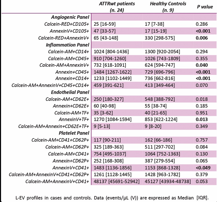
Conclusions: In conclusion, ATTRwt patients display a L-EV profile characterized by endothelial activation and inflammation. Platelet- and endothelial-derived L-EVs were similar between cases and controls. Our findings support EVs as potential biomarkers of vascular and immune dysfunction in the studied population though their clinical role remains to be elucidated.
Downloads
Citations


How to Cite

This work is licensed under a Creative Commons Attribution-NonCommercial 4.0 International License.