CS07 | Endothelial dysfunction and impaired angiogenesis in Hermansky-Pudlak syndrome (HPS) type 1: studies with endothelial colony forming cells
F. Tondi1, G. Purgatorio1, C. Sgromo2, E. Giglio1, E. Petito1, E. Falcinelli1, G. Guglielmini1, A. Follenzi2, P. Gresele1, L. Bury1*, C. Olgasi3* | *Shared senior authorship; 1Department of Medicine and Surgery, Section of Internal and Cardiovascular Medicine, University of Perugia; 2Department of Health Sciences, School of Medicine, University of Piemonte Orientale, Novara; 3Department of Translational Medicine, School of Medicine, University of Piemonte Orientale, Novara, Italy
All claims expressed in this article are solely those of the authors and do not necessarily represent those of their affiliated organizations, or those of the publisher, the editors and the reviewers. Any product that may be evaluated in this article or claim that may be made by its manufacturer is not guaranteed or endorsed by the publisher.
Authors
Background and Aims: Hermansky-Pudlak syndrome (HPS) is an inherited multisystemic disorder caused by a defect of lysosome-related organelles (LRO), like delta granules in platelets, melanosomes in melanocytes and Weibel Palade Bodies (WPB) in endothelial cells (EC). HPS is characterized by oculocutaneous albinism with nystagmus, bleeding diathesis and, in some cases, extra-hematological complications such as pulmonary fibrosis, granulomatous colitis and immunodeficiency. Eleven genes have been identified as causative for HPS in humans. The most severe subtype of HPS is HPS-1, in which variants affect the HPS1 protein. The mechanisms involved in the pathogenesis of HPS have already been investigated in melanosomes, alveolar epithelial cells and platelet delta granules but little is known about EC function in HPS, except for one study showing impaired maturation of WPB in endothelial colony forming cells (ECFC) isolated from an HPS-2 patient. Our aim was to investigate endothelial function, WPB secretion and angiogenesis of ECFC isolated from an HPS-1 patient.
Methods: ECFC were isolated from peripheral blood of a HPS1 patient and from healthy controls. WPB were quantified by immunofluorescence (IF). Von Willebrand Factor (VWF) content and release was evaluated by enzyme-linked immunosorbent assay (ELISA), angiopoietin 2 (Ang-2) secretion was assessed by western blotting (WB) under resting conditions or upon stimulation with histamine (100 μM). Migration was assessed by the scratch-wound healing assay, and angiogenesis by the Matrigel-based tube formation assay. iPSCs were isolated from peripheral blood CD34+ cells of a HPS1 patient and were characterized for exogenous markers, embryoid bodies generation and three germ layers characterization.
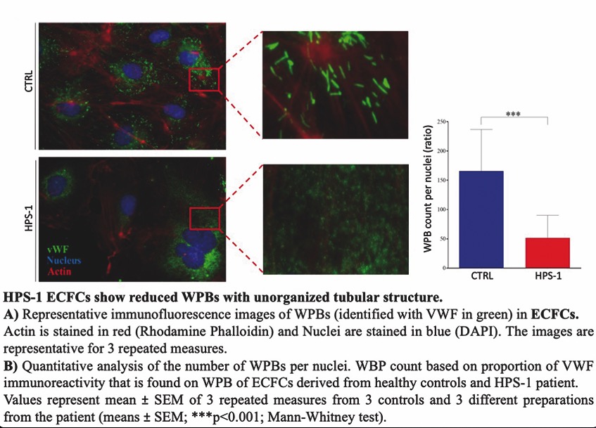
Results: WPBs have the typical elongated morphology in control ECFC, but they were significantly reduced and with unorganized structure in HPS-1 ECFC (Figure 1). Secretion of VWF was significantly reduced in HPS-1 as shown by increased levels of intracellular VWF and decreased presence of VWF in supernatants upon stimulation with histamine in HPS-1 respect to control ECFCs. Moreover, HPS-1 ECFCs showed a significantly decrease in Ang-2 secretion under stimulation with histamine compared to healthy control ECFCs. In addition, the scratch assay showed a significantly higher residual area in HPS1 (65.1±4.7% vs. 33.1±4.3%; p<0.05), showing defective EC migration. Finally, HPS1 ECFC formed a reduced total tube number (113±2.1 vs. 153±2.7, p<0.001) with shorter length (11.2±0.3mm vs. 17±0.7mm, p<0.001) and lesser branches (57.4±2.2 vs. 75.2±2.9, p<0.001) compared to controls, showing defective angiogenesis. CD34+ cells from the HPS-1 patient were successfully reprogrammed into iPSCs. Obtained colonies showed a typical embryonic stem cells-like morphology, 3 clones were selected and were positive for the alkaline phosphatase staining and for the stem cell markers Oct4, Sox2, Klf4, Ssea3 and Tra 1-60. Finally, they were able to mimic embryogenesis at the level of the gastrula by generating embryoid bodies.
Conclusions: Our results show that HPS1 mutation alters the structure of WPB and the expression of proteins stored in these organelles in endothelial cells with associated defective EC migration and angiogenetic activity. Endothelial dysfunction may play a role in the HPS1 bleeding phenotype. A future step of our research will be to exploit iPSCs to test a gene therapy approach to diseased cells.
Downloads
Citations


How to Cite

This work is licensed under a Creative Commons Attribution-NonCommercial 4.0 International License.
Similar Articles
- Guest Editor: Valerio De Stefano, 29th National Conference of the Italian Society for the Study of Hemostasis and Thrombosis, 2025 , Bleeding, Thrombosis and Vascular Biology: Vol. 4 No. s1 (2025)
- CS08 | A combination of two rapid immunoassays improves HIT diagnosis , Bleeding, Thrombosis and Vascular Biology: Vol. 4 No. s1 (2025)
- Silvia Linari, Renato Marino, Marzia Leotta, Antonio Coppola, Patrizia Di Gregorio, Augusto Bramante Federici, Flora Peyvandi, Cristina Santoro, Ezio Zanon, Rita Carlotta Santoro, The landscape of rare coagulation factor deficiency management in Italy: a national hemophilia center survey , Bleeding, Thrombosis and Vascular Biology: Vol. 4 No. 3 (2025)
- Roger Lijnen, Désiré Collen, The key to fibrinolysis and thrombolysis , Bleeding, Thrombosis and Vascular Biology: Vol. 4 No. 3 (2025)
- Bianca Clerici, Mariangela Scavone, Gian Marco Podda, Recent advances in classic heparin-induced thrombocytopenia (HIT), autoimmune HIT, spontaneous HIT, and vaccine-induced immune thrombotic thrombocytopenia , Bleeding, Thrombosis and Vascular Biology: Vol. 3 No. 2 (2024)
- CO44 | First interlaboratory validation workshop of immunofluorescence microscopy on the peripheral blood smear for recognizing patients with inherited platelet disorders , Bleeding, Thrombosis and Vascular Biology: Vol. 4 No. s1 (2025)
- Luca Puccetti, Vincenzo Sammartano, Federico Caroni, Margherita Malchiodi, Paola Calzoni, Eleonora Franceschini, Lucrezia Galasso, Monica Bocchia, Safety of COVID-19 mRNA vaccination in patients with history of acquired hemophilia A: a case series , Bleeding, Thrombosis and Vascular Biology: Vol. 1 No. 3 (2022)
- PO03 | Enlarged platelets with reduced GP IB/IX can indicate disorders other than Bernard-Soulier syndrome , Bleeding, Thrombosis and Vascular Biology: Vol. 4 No. s1 (2025)
- Marcello Di Nisio, Matteo Candeloro, Nicola Potere, Ettore Porreca, Jeffrey I. Weitz, Factor XI inhibitors: a new option for the prevention and treatment of cancer-associated thrombosis , Bleeding, Thrombosis and Vascular Biology: Vol. 3 No. s1 (2024)
- Renato Marino, Silvia Linari, Marzia Leotta, Antonio Coppola, Chiara Biasoli, Maria Elisa Mancuso, Flora Peyvandi, Cristina Santoro, Ezio Zanon, Rita Carlotta Santoro, Diagnostic and management practices for inherited fibrinogen disorders: a nationwide survey of Italian Hemophilia Treatment Centers , Bleeding, Thrombosis and Vascular Biology: Vol. 4 No. 3 (2025)
You may also start an advanced similarity search for this article.